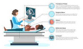
Ultrasound is a scanning technique that uses high frequency sound waves
As a scan is done. The pictures projecte onto screen to create a moving image. A good example of this is the scanning done during pregnancy that can show images of your unborn baby.
Types of ultrasound
There are 3 main types of ultrasound scans:
- External scan – the ultrasound probe is move over the skin on the outside of your body. This is the most common type of ultrasound scan.
- Internal scan – the probe is inserte into your vagina or rectum get a better view of nearby parts.
- Endoscopic scan – the probe is attached to tube and pass further into the body. To examine deeper parts of your body.
Ultrasound scans can use to diagnose conditions. To assess the size and function of your organs and to assess and monitor your baby before they’re born. They can also use to guide a doctor or other healthcare provider as they carry out some procedures or surgeries.
Common areas that can scanne are your:
- abdomen (tummy/puku)
- breast (see below)
- kidneys
- liver
- pelvis – in women this commonly checks your ovaries, bladder and uterus wall thickness. In men it can use to check your prostate, bladder and testicles
- heart – this by ultrasound scan known as echocardiogram or ECHO
- arteries and veins – to check the blood flow in your vessels and check for clots
- uterus and baby during pregnancy
- joints – ultrasound of joints can use to assess the ligaments, tendons and fluid in your joint and also check for arthritis.
Ultrasound scans are often used in real เล่นเกมคาสิโน UFABET ทันสมัย ฝากถอนง่าย time by healthcare providers doing procedures. Examples of this may be:
- to guide the taking of a breast biopsy (sample of breast tissue) accurately
- to place a tube for people undergoing hospital based treatments
- to assist with correct positioning of a needle. When fluid is being inject into, or taken out of a joint.